


Up, Close, and Personal: How Microphotography Opened Up New Avenues in Science
Microphotography has transformed scientific research by revealing intricate details of microscopic structures, enabling breakthroughs in biology, medicine, and material science.

Imagine trying to understand how a car works, but you were only allowed to stare at it from a mile away. You wouldn't be able to spot the small spark plug, gears, or the fuel injection system, right?
The living world was the same for scientists for hundreds of years! They were aware of very small things like cells, bacteria, and hidden structures, but couldn't view them clearly, and thus couldn't share their observations either. The technology of microscopy and the camera combined to form microphotography.
This very microphotography idea is one of the most significant inventions in science. It has allowed us to move from theories about how life operates to witnessing it at the cellular level, and this has influenced medicine, engineering, and practically everything else!
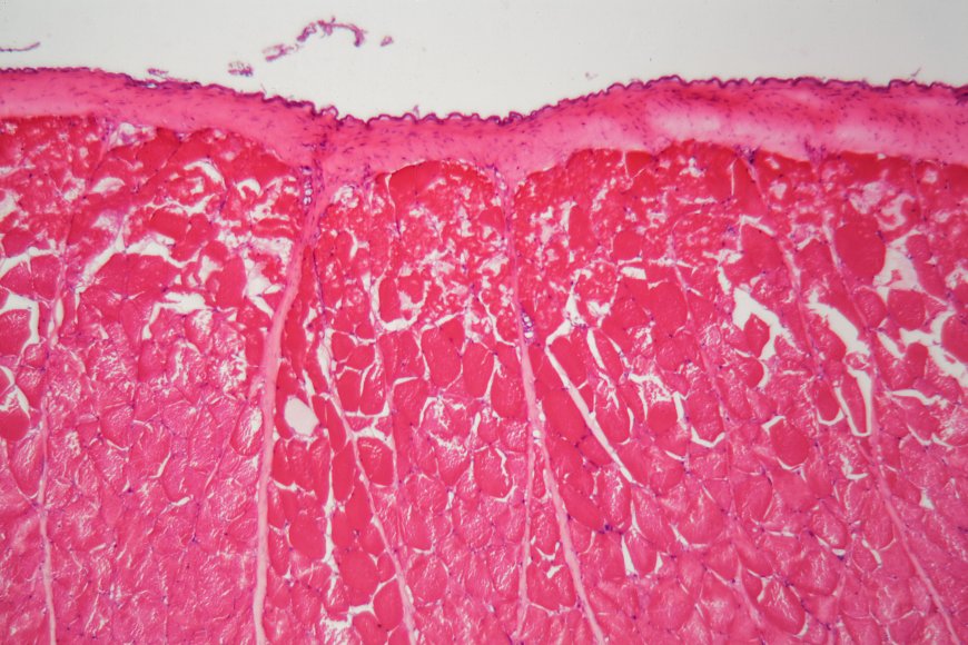
What is Microphotography? (It’s Not Just a Close-Up!)
Before we get into the incredible and interesting science, let's be clear on what we mean.
The definition of microphotography is to take pictures using a microscope.
Generally speaking, photography means reducing a very large object (say, the Eiffel Tower) to a miniature version that fits into the small film or camera sensor.
Microphotography (or Photomicrography) means scaling a tiny, microscopic object (e.g., a single plant cell) to a gigantic size to make the details visible and take a photo of them.
In simpler terms, it is the operation of combining a microscope with a camera to create a micrograph (the output image from the process).
Types of Microphotography
Microphotography is a complex field with various methods. Different microphotography techniques are adopted by scientists based on their observation requirements:
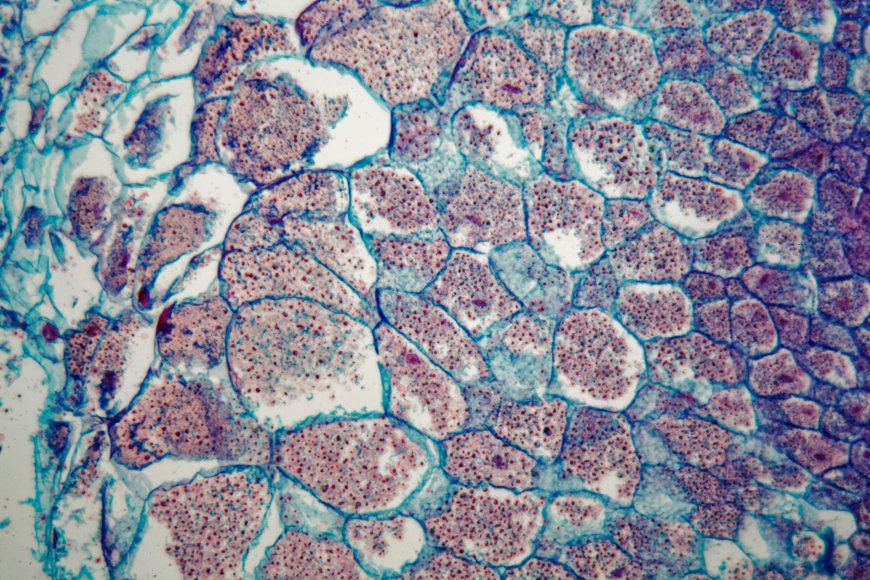
Photomicrography (Light Microscopy)
The prime example. It employs common light for the observation of cells, tissues, and crystals. This is the sort of microscopy that can mostly be found in the bioscience book.
Electron Micrography
Instead of light, a stream of electrons is the source here, which allows for tremendous magnification—so incredibly strong that one can witness minuscule viruses or the interiors of a metallic compound.
To grasp the microphotography concept is to realize its greatest impact: it has made invisible worlds visible, and even more so, it has created a common visual fact arena.
Medicine and Biology: Unlocking the Mysteries of Life
The very first time the significance of microphotography was fully appreciated was in the field of medicine. The period was before drawing was still the only way to document what was observed under the microscope, which was an extremely tedious process and prone to mistakes.
1. Diagnosing Diseases
Microphotography was one of the techniques that made it possible for doctors to get not only top-quality but also lasting pictures of human tissues and cells.
Live Example (Pathology): In the course of the 19th century, Alfred François Donné, among others, was the one who took the first steps to publish photomicrograph atlases. He used this method to first article the likes of platelets and leukemia cells. Today, the same scenario would be when a pathologist takes a micrograph to validate the presence of a disease, for example, cancer, in a small tissue sample. This visual evidence is very much necessary for the accurate diagnosis and thereby selection of the appropriate treatment.
2. Tracking Microorganisms
The images played an essential role in confirming the Germ Theory of Disease.
The Great Impact: After the moment when scientists were able to get bacteria, viruses, and other microorganisms photographed, the sharing of those pictures around the world became possible. This was the proof of the existence of these tiny beings and their role in disease. The understanding became a basis for the adoption of sanitation measures, hygiene practices, and the invention of vaccines and antibiotic drugs that save millions of lives.
3. Understanding Cell Structure
Cell biology owes its entire existence to microphotography. It is not possible to work on something that is not visible! The possibility of taking photographs and monitoring the development and movement of living cells is an absolute must for current research in different fields from developing new drugs to understanding genetics.
Industry and Forensics: Seeing the Smallest Flaws
The applications of microphotography go far beyond biology; they are essential in modern industry and crime-fighting.
1. Quality Control in Manufacturing
The current era has a characteristic that even the tiniest mistakes may lead to major troubles. Microphotography is employed in the inspection of materials to reveal imperfections that are not seen by the unaided eye.
Real-Life Scenario (Semiconductors): The semiconductor chips in your computer and mobile phone consist of extremely small-scale circuits. In the semiconductor sector, such as the microphotography of high-power reflected-light, such as the one used for inspecting the circuits enabled machines during production, are a common practice. The technicians check for minute scratches, fractures, or dust particles that might spoil the chip.
2. Forensic Science and Crime Solving
The tiny world underneath the microscope frequently reveals the most crucial evidence that could lead to a criminal's arrest.
The Role: Forensic scientists resort to microphotography to record and scrutinize minuscule evidence. This is inclusive of photographing the striations (scratch marks) on a discharged bullet for matching it to a particular firearm or looking at the individual hair or fiber samples collected from the scene of the crime. The images created are the permanent records that serve as solid evidence in court.
3. Metallurgy and Materials Science
Scientists have to observe how the new type of steel or metal alloy with high strength and low weight looks internally when they are developed.
The Process: Micrographs of the metal's cross-section are taken to have a look at its grain size and microstructure. The visual analysis aids the scientists in discovering the reasons behind the varying strengths and flexibilities of the materials. This, in turn, contributes to the design of stronger cars, bridges, and airplanes.
The Lasting Importance of Microphotography
The importance of microphotography can be summed up in one idea: it allowed scientists to share the objective truth. Before the camera, a scientific drawing could be disputed. A photograph, however, offered undeniable evidence of the microscopic reality.
Microphotography has come a long way from the earliest plant section images taken by William Henry Fox Talbot in the 1830s to today's gorgeous, colored cell images produced by modern digital cameras. It is still one of the most essential tools in science that not only provides permanent and detailed recording but also enables scientists to teach, compare results globally, and even build on each other's discoveries.
The world we perceive with our naked eyes is only a fraction of reality. Microphotography has enabled us to not only see the invisible, beautiful, and complicated universe but also to resolve some of the most difficult problems in the world!
What's Your Reaction?
 Like
0
Like
0
 Dislike
0
Dislike
0
 Love
0
Love
0
 Funny
0
Funny
0
 Angry
0
Angry
0
 Sad
0
Sad
0
 Wow
0
Wow
0